by Ann Ramsey BS, CERA, APF–I
WHAT IS NAVICULAR?
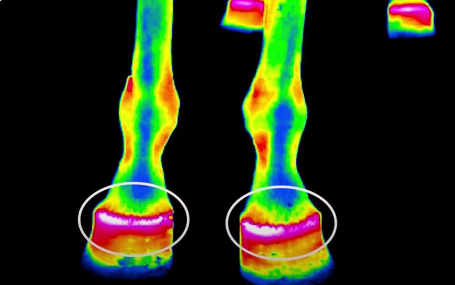
When we say a horse has ‘navicular’ it usually describes a horse with bilateral forelimb lameness. It’s important to understand that ‘navicular’ is really an umbrella term for many different pathologies that can occur at the back of the horse’s foot. Painful heels can be caused by anything from atrophy of the soft tissue that comprises the heel, tendon or ligament lesions, or boney changes to the navicular bone. Critical to the horse’s recovery is to have the veterinarian perform the necessary diagnostics to determine the real source of pain. This should include a full lameness exam and may require nerve blocks, x-rays and MRI assessments.
LANDING TOE FIRST
Despite the multiple sources of chronic caudal heel pain, the movement pattern of affected horses will change in a predictable way; they land toe first instead of heel first. Many horses disguise heel pain this way for years. With this toe first movement pattern horses will appear “sound’ because they can travel evenly when both feet are affected. Unfortunately, the toe first landing can cause further pathology to develop. Dr James Rooney and others have described how a toe first landing can cause damage to the deep digital flexor tendon, and ultimately the navicular bone.
A toe first landing is the first compensation a horse can use to cope with painful heels, and once you can spot that movement pattern its important to stop and evaluate why it’s happening. Your veterinarian and farrier should be called if you notice a horse is landing toe first. The earlier you find and treat the source of pain; the less damage will occur to the caudal foot.
REHABILITATION APPROACHES
As a hoof care provider, I utilize different therapeutic approaches depending on the individual animal’s morphology and disease progression. I have had success using both shod and barefoot methods for rehabilitation. The most important goal is to restore a heel first landing. Everything I use is designed to facilitate a healthy heal first impact. For example, if a horse lacks hoof mass, I add supportive materials like custom orthotics and I choose materials that will help dissipate energy and absorb concussion.

If a horse just simply has some contraction or atrophy of the soft tissue at the back of the foot, a barefoot approach that incorporates judicious use of boots and pads, along with graduated exercise can be very successful as well. The key is to offer a wide range of therapeutic tools and options depending on the individual animals needs.
COLLABORATIVE CARE
Navicular disease treatment hinges on the collaborative care given by your vet and farrier. I work in tandem with a vet to ensure the best diagnostics and ongoing pain management are being utilized. In addition, I use radiographs on a regular basis to shoe or trim lame horses. I request radiographs be taken at the first few shoeings so that I can provide a detailed orthopedic application. Using radiographs is the best way to accomplish a nuanced therapeutic shoeing application, and the quickest way to get the horses trajectory to improve. It’s well worth the cost over the long run.

TRAIN YOUR EYE FOR EARLY DETECTION!
Today vets and farriers have some wonderful tools available that make navicular syndrome less painful for horses and less frightening for horse owners. The best defense against navicular disease is to catch it early. As a horse owner it is critical to learn to spot a toe first landing pattern and do something proactive to intervene. The sooner your horse’s caudal heel pain is resolved the better chance you have at avoiding permanent damage. If your horse has this diagnosis, don’t panic, just get your team assembled.
Pain in the front feet can frequently be resolved, but keep in mind that the front feet are not the only structures affected by the disease process, the neck and thorax and forelimbs of the horse are also affected as they are recruited in compensation. These areas of the body can become painful and require treatment and therapies as well. Once the horse’s front feet are sorted out, I start to use bodywork, targeted exercise and other rehabilitative techniques to further their care.
For more on this topic, see the resources below:
Course
Blog Posts
Neck Bracing and Forelimb Pain (PART 1)
Neck Bracing and Forelimb Pain (PART 2)